Abstract
The synthesis of silver-doped zinc oxide (Ag:ZnO) nanocomposite material was achieved using a simple chemical coprecipitation method, in which 0.2 M zinc chloride and 0.001 M silver nitrate coprecipitated with 25% ammonia solution by pulse mode dispersion using ultrasonicator. The obtained silvery white precipitate was dried overnight at 110°C in hot air oven, and the powder was collected. The resulted Ag:ZnO nanocomposite was structurally and optically characterized using various techniques. The X-ray diffraction (XRD) pattern clearly showed the presence of crystalline Ag:ZnO particles. Further, UV-Vis spectrophotometer and fourier transform infrared spectroscopy (FT-IR) results showed the presence of Ag:ZnO nanocomposite at specific wavelengths. The scanning electron microscopy (SEM) and transmission electron microscopy (TEM) analysis confirm that the synthesized Ag:ZnO nanocomposite material was truncated nanorod in shape and has 48 to 226 nm size in diameter.
1. Introduction
Zinc oxide (ZnO) is an important industrial material, because it has an inorganic and semiconducting material with inherent properties that share its structure as wurtzite [1]. ZnO nanocomposite has attracted interest because of its optical properties. These particles are transparent to visible light, but they absorb UV-light. ZnO has attracted intensive research effort for its unique properties and versatile applications in transparent electronics, chemical sensors, and spin electronics [2–8]. A variety of metal oxides like zinc oxide, titanium dioxide (TiO2), and silicon dioxide (SiO2) and different techniques such as chemical coprecipitation [9, 10], sol-gel process [11, 12] chemical vapour deposition [13], thermal decomposition [14, 15], hydrothermal synthesis [16, 17], solid-state reaction [18], spray pyrolysis [19], vapour-liquid-solid method [20], and microemulsion precipitation [21–23] have been used so far. Hingorani et al. also reported the synthesis of ZnO nanoparticles (NPs) and it was the first study using reverse microemulsion in the early 1990s [24, 25].
Synthesis process of Ag:ZnO nanocomposite plays a key role in governing size, morphology, and its properties of nanocomposite through dispersion method (pulse mode), using ultrasonication [26]. The main objective of synthesis is to produce particles only in nanorod structure for various toxicity and biological application studies. There are different doping agents like P [27], N [28], As [29], Li [30], Sb [31–34], and Ag [35]. Among these, we have taken Ag as doping agent. Because the nature of Ag ions is simple link matrices, their behaviour to surface states in nanomaterials where the surfaces of are becomes prime importance as the size decreases. The sonochemical synthesis of nanomaterial is extensively used to generate novel materials with different properties, because this method can produce similar truncated nanorods with smaller size than those reported by other methods [36–38]. The advantages of chemical coprecipitation method through sol-gel process for preparing nanocomposite materials are to get small average particle size and narrow size distribution, homogeneity, purity, and more possibility to obtain nanorods.
2. Materials and Methods
All chemicals used in this study (zinc chloride 98%, silver nitrate 99.8%, and ammonia 25%) were AR grade purchased from HiMedia, Mumbai, India. All reagents were prepared by using deionised water (18 MΩ resistivity).
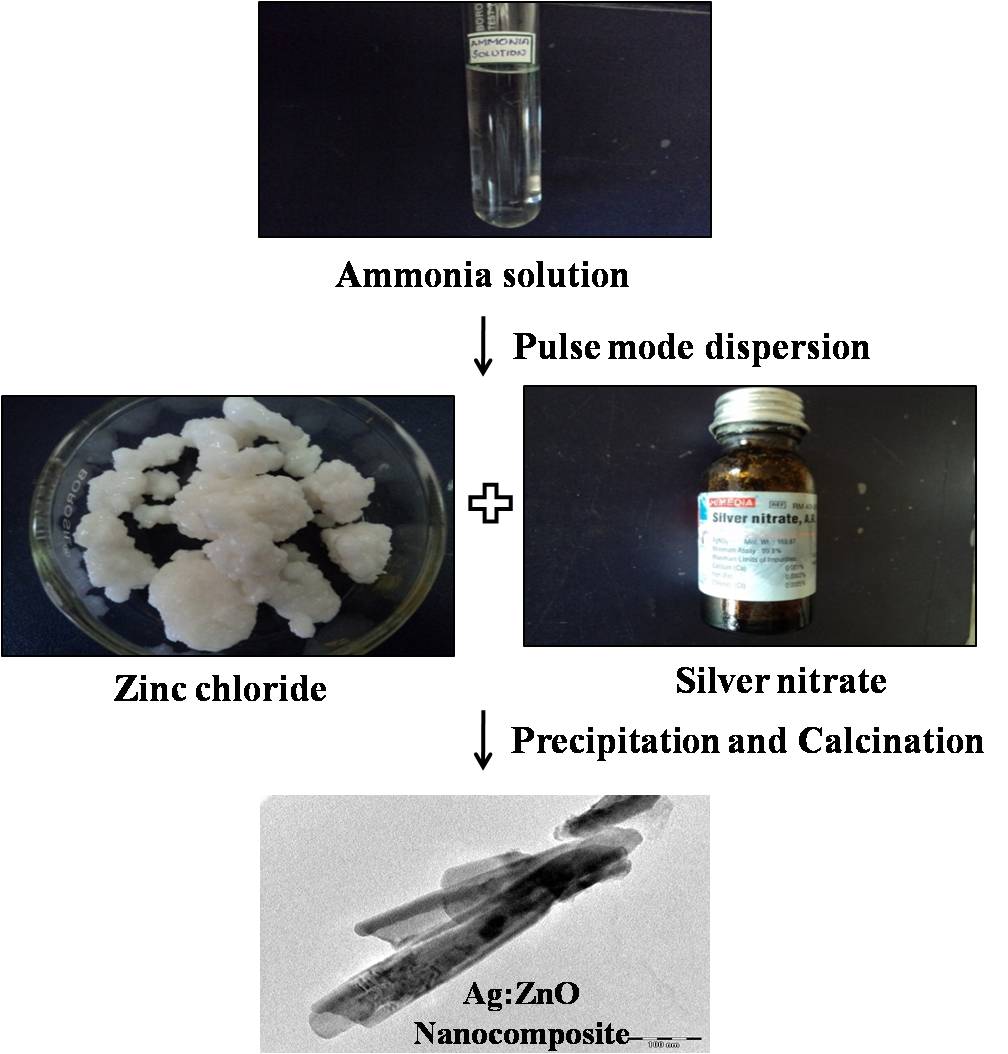
2.1. Synthesis of Ag-Doped ZnO Nanocomposite
0.2 M zinc chloride (ZnCl2) and 0.001 M silver nitrate (AgNO3) were mixed to 50 mL of distilled water. A 2 mL of 25% ammonia solution was added dropwise until precipitation occurred, and then further 10 drops of ammonia solution (NH4OH) were added to make the solution clear. Pulse mode sonication (PS) was operated at 112.5 W, and the frequency of the sonicator were maintained at 20 kHz ± 50 Hz. PS takes place for one second then stops for one second, and the total process took two hours to get precipitates. The initial pH of the solution was 10.2, whereas at the end, the pH was 8.2 and the solution became clear silvery white, and gradually solid suspension is settled. The formed precipitate was washed several times with deionised water and acetone followed by centrifugation at 3500 rpm for the complete elimination of chloride. The precipitate was then dried at room temperature and kept overnight inside an oven at 110°C in atmospheric pressure to obtain dry powder.
Ammonia solution (NH4OH) was used as precipitating agent during synthesis between zinc chloride (ZnCl2) and silver nitrate (AgNO3) with water (H2O) to form Zn(OH)2, AgOH, NH4Cl, NH4NO3, and H2O. The product was washed with deionised water for several times to form Zn(OH)2, AgOH, and H2O, then again washed with acetone by centrifuging in order to form Zn(OH)2 and 2AgOH. On heating at 110°C the product obtained was pure ZnO-doped Ag2O like (Zn–O–Ag) in the precursor. Through the solvothermal process, the precursor reduced by acetone gradually and Zn–O–Ag bond formation between Ag-doped ZnO nanocomposite materials, which has been explained as
2.2. Characterization of Ag:ZnO Nanocomposite Material
Several techniques were used to characterize Ag:ZnO nanocomposite material like UV-Visible spectroscopy, XRD and FT-IR. The crystalline nature of nanocomposite was characterized by XRD, using PANalytical Xpert-Pro diffractometer equipped with Cu Kα1 radiation (). Morphology of the sample was studied by using SEM (Model FEG-Nova 600 Nano SEM). The UV-Vis spectra were recorded with multimode microplate reader (Synergy HT) in the wavelength range between 300 and 800 nm. An FT-IR spectrum was recorded using Perkin-Elmer instrument (model spectrum RX1). Further Ag:ZnO nanocomposite material was characterized by using various techniques like DLS and TEM.
3. Results and Discussion
This work reports that the synthesis of Ag-doped ZnO nanocomposite material through chemical coprecipitation method overpoweringly achieves the synthesis through pulse mode dispersion by ultrasonication. In this study, it was clearly observed that the catalyst ammonia plays a primary role in forming the shape of a particle during the synthesis process. Moreover, the shape of the nanocomposite changed to rod from spheres when it was doped with silver ions, and it was an interesting morphology observed through TEM analysis. Most of the reports were about silver and gold nanocomposite [39, 40], but only a few data were available for doping of wide band semiconductor like ZnO with silver ions. The comprehensive structural studies are not available, and the methods of preparation of doped NPs are still evolving [41, 42]. In the synthesis method, pH also plays an important role in the formation of diverse morphology [43].
3.1. X-Ray Diffraction Study
The average crystallite size can be determined through Full Width at Half Maximum (FWHM) of X-ray diffraction peak by using Debye-Scherer’s equation as where is grain size (nm), is wavelength of the XRD used, is FWHM, and is Bragg’s angle. X-ray diffractogram was analyzed to obtain information about various crystalline aspects of Ag:ZnO nanocomposite material. The X-ray diffraction patterns of Ag:ZnO nanocomposite material are wurtzite hexagonal phase [44] and were shown in Figure 1 which indicates the well-indexed XRD peaks corresponding to the planes (100), (002), and (101), JCPDS no. 89-1397. The peak (103) indicates the presence of silver (Ag), and no other impurities were observed (the standard file of JCPDS no. 89-3722). These results indicate that the products consisted of pure phases. Furthermore, the diffraction peaks were more intensive and narrower, implying a good crystalline nature of Ag:ZnO nanocomposite products. The size range of Ag:ZnO nanocomposite material was from 26 nm to 136 nm in diameter according to Debye-Scherer’s equation.

3.2. Fourier Transform Infrared Spectroscopy Study
Synthesized Ag:ZnO nanocomposite material was analysed by FT-IR in the range from 400 to 4000 cm−1 at room temperature (Figure 2). The FT-IR spectrum of the Ag:ZnO nanocomposite material contains several bands with remarkable features. The bands between 400 and 750 cm−1 correlated to metal oxide bond (ZnO) [45–47]. Bands around 900–1500 cm−1 are due to the oxygen stretching and bending frequency [48, 49]. The spectral band at 463.01 cm−1 and band at 723.96 cm−1 clearly show the presence of ZnO and Ag ions approximately equal to those reported in the literature [50]. On doping, stronger and wider absorption were bands observed in the region ~723.96 cm−1 due to the organic capping of silver [51]. Bands at 1394.82 cm−1 and 1507.07 cm−1 correspond to C=O and O–H bending vibrations, respectively. The presence of weak band near 1604.07 cm−1 is assigned to H–O–H bending may due to the adsorption of moisture. Bands at 3465.60 cm−1 indicate the presence of OH group. It is evident from the FTIR data that the Zn–O vibrational mode was more prominently observed, and this clearly concludes a strong doping between Ag and ZnO nanocomposite materials [52].

3.3. UV-Visible Absorption Study
The UV-Vis absorption spectra (Figure 3) of Ag:ZnO nanocomposite material were recorded at room temperature. The synthesized nanocomposites were dispersed in ethanol with the concentration of 0.1% water, and the solution was used to record UV-Vis spectra at wavelength range between 300 and 600 nm. The spectra reveal a characteristic absorption peak of Ag:ZnO nanocomposite material at 360 nm [53, 54]. The absorbance increases in the higher wavelength side indicating the role of nano Ag particles in ZnO. More numbers of Ag are dispersed into ZnO, and these metallic particles rest on the surface of ZnO nanocomposite making the surface area increased and showing plasmonic resonance peak in the UV-Vis spectra. This is an important method in which Ag:ZnO composite nanorod has been shown and elevated by UV absorption. This high absorbance of the Ag:ZnO composite nanorods has an important implication for various industrial applications.

3.4. DLS Study
Malvern particle size analyzer (Model, Micro-P) was used to measure the size of synthesized Ag:ZnO nanocomposite materials. The average size obtained by the DLS distribution plot was around 60 to 300 nm, in this 163 nm particles were more (Figure 4). The dimension of Ag-doped zinc oxide averages in the form of an equivalent sphere and hence does not give the distribution in the range of lengths or widths of the nanorods. The average sizes from DLS result that the particle also coincide with SEM and TEM results [55].

3.5. SEM and TEM Analyses of the Ag:ZnO Nanocomposite Materials
The SEM uses a focused beam of higher energy electrons to generate a variety of signals at the surface of solid specimens. The signals that derive from electron sample interactions reveal information about the sample including external morphology. When samples were viewed under SEM, the truncated nanorod morphology with irregular size was observed, which was randomly orientated. The diameter and length of the Ag-doped zinc oxide nanorod were found, approximately 54 to 226 nm diameters and from 601 to 1420 nm length with one-dimension were segregated (Figures 5(a) and 5(b)).

(a)

(b)
TEM characterization was carried out in diffraction and imaging modes by using FEI-TECNAI G2-20 working at 200 KV. Images of Ag:ZnO NPs shown in Figures 6(a) and 6(b) are monodisperse, and the selected area diffraction (SAED) pattern apparently showed clear truncated nanorod structure of the nanocomposite. From the TEM micrograph, the diameter of Ag-doped zinc oxide was found to be approximately 48 to 126 nm and the length was 232 to 1255 nm with one-dimension of nanorod. The doping of silver with zinc oxide nanocomposite material changes spherical ZnO to rod shape Ag:ZnO particles. The entire particle sizes results obtained were almost the same in XRD, DLS, SEM, and TEM when comparing with one another. Finally, The shape and size of the Ag:ZnO nanocomposite was confirmed by the comprehensible techniques such as SEM and TEM.

(a)

(b)
4. Conclusions
From the above presented experimental results and discussion, it is clear that the synthesis through ultrasonication method gives the following conclusions.(a)Chemical Coprecipitation is an easy and simple method that played an efficient task in the synthesis of Ag:ZnO nanocomposite material when compared with other methods.(b)In pulse mode synthesis, precursor and reducing agent are main reasons for the morphological changes during synthesis.(c)Addition of Ag+ ions during dispersion (Pulse mode) through ultrasonication increases high crystalline nature and change in morphology. (d)Ag+ ion rests upon the surface of the zinc oxide at some stage to form truncated nanorod morphology.(e)Temperature, time, and alteration of initial and final pH also play prominent role in forming different size and morphological evolution of Ag-doped zinc oxide nanocomposite (Ag:ZnO).
Finally, it was concluded that the several unique features of synthesized Ag:ZnO were observed with the deserved special comments. The generality and simplicity for the synthesis of Ag-doped ZnO nanocomposite material are involved in trouble-free steps. Several researchers have recently reported the chemical coprecipitation synthesis of nanocomposite, nanowires, and nanorods [56–59]. However, our chemical coprecipitation method allows the synthesis of nanorods in easy way where the dispersion method by ultrasonicator plays novelty in the formation of nanorod Ag:ZnO.
Acknowledgments
The authors owe sincere thanks to UGC and UGC-RFSMS for providing financial support and MoE&F for providing instrumentation facility. The authors thank Dr. N. P. Lalla UGC-DAE CSR Indore for TEM measurement that is highly appreciable and commendable. Sincere thanks are to Department of Chemistry and Physics, Bharathidasan University, Tiruchirappalli, for their valuable suggestions and comments.
Supplementary Materials
The Synthesis of Silver-doped Zinc oxide (Ag:ZnO) Nanocomposite material was achieved using a simple chemical co-precipitation method. In which 0.2 M zinc chloride and 0.001 M silver nitrate co-precipitated with 25% ammonia solution by pulse mode dispersion using ultrasonicator. The obtained slivery white precipitate dried overnight at 110ºC in hot air oven and collected the powder. The resulted Ag:ZnO nanocomposite was structurally and optically characterized using various techniques. The X-ray Diffraction (XRD) pattern clearly showed the presence of crystalline Ag:ZnO particles. Further, UV-Vis spectrophotometer and Fourier Transform Infrared Spectroscopy (FT-IR) results showed the presence of Ag:ZnO nanocomposite at specific wavelengths. The Scanning Electron Microscopy (SEM) and Transmission electron Microscopy (TEM) analysis conforms that the synthesized Ag:ZnO Nanocomposite material was truncated nanorod in shape and has 48 to 226 nm size in diameter.